OCT
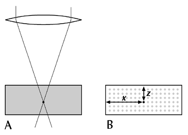
Optical coherence tomography (OCT) is a very different technique to OPT(!) It can be described as an optical equivalent of ultrasound. Light is directed into a specimen, and the intensity and time-of-flight of reflected photons is calculated using low-coherence interferometry. The technique therefore samples the reflectiveness of the specimen along a line parallel to the optical axis. This line is essentially a 1-dimensional section through the specimen, and the technique is thus another example of section tomography.


